Category Archives: Biochemistry and Biophysics

Proteins run the show (except when they unfold and cause cataracts)

Your eye lenses host one of the highest concentrated proteins in your entire body. The protein under investigation is called crystallin and the investigator is called Heather Forsythe.

Heather is a 4th year PhD candidate working with Dr. Elisar Barbar in the Department of Biochemistry and Biophysics. The Barbar lab conducts work in structural biology and biophysics. Specifically, they are trying to understand molecular processes that dictate protein networks involving disordered proteins and disordered protein regions. To do this work, the lab uses a technique called nuclear magnetic resonance (NMR). NMR is essentially the same technology as an MRI, the big difference being the scale at which these two technologies measure. MRIs are for big things (like a human body) whereas NMR instruments are for tiny things (like the bonds between amino acids which are the building blocks of proteins). Heather employed OSU’s NMR facility (which has an 800 megahertz magnet and is on the higher end of the NMR magnetic field strength range) to investigate what the eye lens protein crystallin has to do with cataracts.

Your eye completely forms before birth, and the lens of the eye that helps us see is made of a protein called crystallin. This protein is essential to the structure and function of the eye, but it cannot be regenerated by the body so whatever you have at birth is all you will ever have. However, in the eye lens of someone affected by cataracts, the crystallin proteins become unfolded and then aggregate together. They stack on top of each other in a way that they are not supposed to. A person with cataracts will suffer from blurry vision, almost like you’re looking through a frosty or fogged-up window. While the surgery to fix cataracts (which basically takes out the old lens and puts in a new, artificial one) is pretty straight-forward and not very invasive, it isn’t easily accessible or affordable to a lot of people all over the world. Cataracts is attributed to causing ~50% of blindness worldwide, likely due to the fact that not everyone is able to take advantage of the simple surgery to fix it. Therefore, understanding the molecular, atomic basis of how cataracts happens could result in more accessible treatments (say a type of eye drop) for it worldwide.

This is where Heather comes in. There are different types of crystallin proteins and Heather zeroed in on one of them – gamma-S. Gamma-S is one of the most highly conserved proteins (meaning it hasn’t changed much over a long time) among all mammals, which tells us that it’s super important for it to remain just the way it is. Gamma-S makes up the eye lens by stacking on top of itself, making a brick wall of sorts ensuring that the eye lens retains its structure. However, research prior to Heather’s found that with increased age there is an increase in a modification called deamidation, which occurs in the unstructured loops of the gamma-S protein. Deamidation is a pretty minor change and is common in proteins all over the body, however in the eye lens if too much of it happens it no longer is a minor issue since it starts to disrupt the structure and protein-protein interactions of the eye lens. Heather’s collaborators at Oregon Health Sciences University found that there are two sites on the gamma-S protein (sites 14 and 76) where these deamidation events increase the most in cataracts-stricken eyes. It’s been known for a while that this deamidation is associated with cataracts however we never knew why it is associated with cataract formation because the changes caused by this modification were seemingly minor. This is how the Barbar Lab, and Heather specifically, became connected to this work since they specialize in studying unstructured proteins and protein regions, such as the loops present in gamma-S.
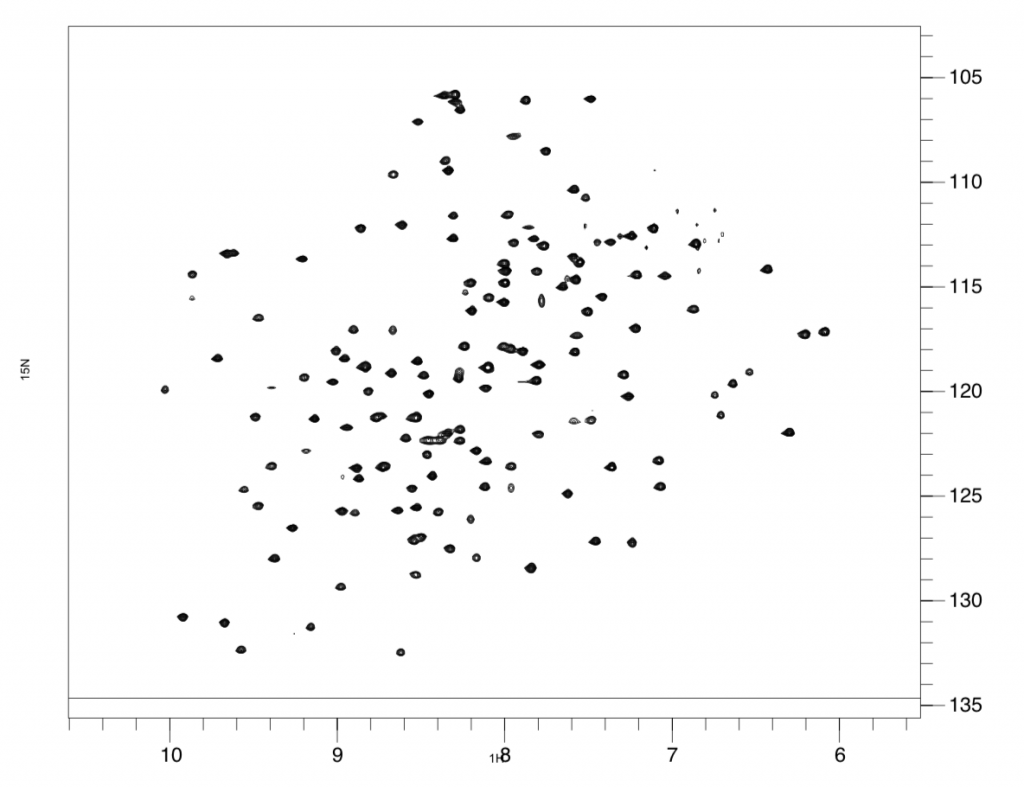
An example of an “1H(x-axis) 15N(y-axis) HSQC” spectra, aka, the fingerprint of a protein. This spectra is of WT gamma-S crystallin.

These deamidation changes are mimicked in the lab by creating two different mutants of the gamma-S protein’s DNA. Heather then compared the two mutants with the normal DNA by putting them through a series of experiments using the trusty NMR. The NMR is basically a large magnet that can make use of the magnetic fields around an atom’s nucleus to determine protein structure and motions. When Heather puts a protein sample into the NMR, the spins of the atomic nuclei will either align with or against the magnetic field of the NMR’s magnet. The NMR spits out spectra, which look like a square with lots of polka dots. This is essentially the fingerprint of the protein, unique to each one and extremely replicable. Heather can analyze this protein fingerprint since the different polka dots represent different amino acids in the gamma-s protein. Heather can compare spectra of the two mutants to the spectra of the normal protein to see whether any of the dots have moved, which would signal a change in the position of the amino acids.

After running experiments which measure protein motions at various timescales, from days to picoseconds, Heather discovered significant changes in protein dynamics when either site 14 or 76 was deamidated, however at different timescales. What this discovery means is that if both of these mutations are associated with cataracts and they are changing the same regions of the gamma-S protein, then these regions are likely central to changes resulting in cataracts. Therefore, research could be directed to target these regions to perhaps come up with solution to prevent and/or solve cataracts in a non-surgical way. The results of Heather’s study were recently published in Biochemistry.
Heather with her dog Piper.

Heather is from Arkansas where she completed her high school and undergraduate education. Living in a single-parent, non-academic home at this time, it took Heather a long time to figure out how to navigate the scientific and college-application scene, as well as even coming to the realization that science was something she was good at and could pursue. Despite receiving scholarships for college, she still had to work multiple jobs while in high school and college to have enough money for car-payments and gas to get to extra-curricular activities and volunteer jobs in the science field; things critical for graduate school applications. As a result, Heather is a strong advocate for inclusivity, striving to make things like science and college in general more accessible to low-income and diverse students. Heather’s decision to leave Arkansas and come to the PNW was inspired by advice she received from her undergraduate advisor who told her “not to go anywhere where you wouldn’t want to live. You will learn to love research, whatever it ends up being, but if you live in an environment that you don’t find fulfilling, then you are going to suffocate.”. Following this advice has lead Heather to where she is now – the senior in her lab where she has become a mentor to undergraduates, makes Twitter-famous Tik Tok videos (see below), goes on adventures with her dog Piper, and publishes cutting edge structural biology research.

Heather and her undergraduate mentee performing The Git Up in the lab.

To learn more you can check out the Barbar Lab website and Twitter page.

To hear more about Heather’s research, tune in on Sunday, September 29th at 7 PM on KBVR 88.7 FM, live stream the show at http://www.orangemedianetwork.com/kbvr_fm/, or download our podcast on iTunes!

Micro structures and macro support

Our guest this week, Shauna Otto from the Department of Biochemistry and Biophysics, is a member of the lab of Dr. Colin Johnson. The focus of the Johnson lab is a group of proteins called ferlins. The ferlin family of proteins have many different functions, and many are involved in the fusion of vesicles to cell membranes in a process called, “exocytosis.” Another example is the protein otoferlin which fuses vesicles carrying neurotransmitters to the cell membrane of neurons in the inner ear that play a crucial role in hearing. See more about otoferlin from past guests from the Johnson lab, Murugesh Padmanarayana and Nicole Hams.

Shauna loading a sample for Cobalt-60 irradiation at the Notre Dame Radiation Laboratory.

Shauna studies dysferlin, another ferlin protein, which helps mend membrane tears in muscle cells. Mutations in the dysferlin gene lead to Muscular Dystrophy II. Through her work, Shauna has characterized portions or “domains” of the large dysferlin protein via Nuclear Magnetic Resonance (NMR). NMR is a process by which the magnetic field around the nuclei of atoms in a protein domain are excited, and by recording the magnitude of that disruption, Shauna can learn the structure of the domain. Her focus domain putatively binds other proteins that join dysferlin in a protein complex that initiates muscle cell membrane repair. However, the mechanism by which dysferlin bind repair proteins is unclear. Through her explorations with dysferlin, Shauna has found that an increase in Calcium leads to the stabilization of the dysferlin domains that might initiate repair. Right now, it is unclear if this stabilization initiates muscle cell repair, but if it does the next question is how and when such stabilization occurs.

Shauna and husband (Kris Hill) backpacking in Yosemite

Shauna’s academic journey was wrought with hardship, and we are grateful to her for being willing to share her story with us on air. Shauna started undergraduate with an interest in marine biology, but found that college is cost prohibitive. After a two year break, she went back to University of California Long Beach to major in Chemical Engineering, but finally landed on biochemistry. She had a knack for chemistry and loved solving complex puzzles in cellular biology through the lens of protein interactions and biochemical pathways. She began undergraduate research, but her work took a turn as she struggled with homelessness. Homelessness is a growing problem for college students, and has prompted bills targeting the problem of home insecurity for students in California and Washington. However, for Shauna, homelessness was not discussed among fellow students and officials when she attended school. Rather, instead of resources to alleviate her financial hardship, she was met with policy allowances such as permission to sleep in her research lab.

Shauna and her daughter in a bookshop.

Since beginning her PhD at OSU, Shauna has found support here on campus from mentors and her department who have listened and replied with support in the form of University Resources and Services to help her succeed academically, financially, and in personal wellness. Given her past, Shauna now knows the questions to ask about support when seeking the next job, and she is a resource for undergraduates and graduate students who are going through similar life experience.

Hear more about Shauna’s research and personal story this Sunday June 2, 2019 at 7 pm on KBVR Corvallis 88.7FM. Stream the show live or catch the episode as a podcast in the coming weeks.

Treating the Cancer Treatment: an Investigation into a Chemotherapy drug’s Toxic Product

One of the most difficult hurdles in cancer treatment development is designing a drug that can distinguish between a person’s healthy cells and cancer cells. Cancerous cells take advantage of the body’s already present machinery and biochemical processes, so when we target these processes to kill cancer cells, normal, healthy cells are also destroyed directly or through downstream effects of the drug. The trick to cancer treatment then is to design a drug that kills cancer cells faster than it harms healthy cells. To this end, efforts are being made to understand the finer details that differentiate the anti-cancer effects of a drug from its harmful effects on the individual. This is where the research of Dan Breysse comes in.

Dan a third-year master’s student working with Dr. Gary Merrill in the department of Biochemistry and Biophysics. Dan’s research focuses on a common chemotherapy drug, doxorubicin. Doxorubicin has been researched and prescribed for about 40 years and has been used as a template over the years for many other new drug derivatives. This ubiquitous drug can treat many types of cancer but the amount that can be administered is limited by its toxic effect on the individual. Nicknamed “the red death,” doxorubicin is digested and ultimately converted to doxorubicinol, which in high doses can cause severe and fatal heart problems. However, hope lies in the knowledge that doxorubicinol generation is not related to the drug’s ability to kill cancer cells. These mechanisms appear to be separate, meaning that there is potential to prevent the heart problems, while keeping the anti-cancer process active.

Cancer cells replicate and build more cellular machinery at a much faster rate than the majority of healthy cells. Doxorubicin is more toxic to fast-replicating cancer cells because its mechanism involves attacking the cells at the DNA level. Dividing cells need to copy DNA, so this aspect of doxorubicin harms dividing cells faster than non-dividing cells. It is common for chemotherapy drugs to target processes more detrimental to rapidly dividing cells which is why hair loss is often associated with cancer treatment.

Separately, doxorubicin’s heart toxicity appears to be regulated at the protein level rather than at the DNA level. Doxorubicin is converted into doxorubicinol by an unknown enzyme or group of enzymes. Enzymes are specialized proteins in the cell that help speed up reactions, and if this enzyme is blocked, the reaction won’t occur. For example, an enzyme called “lactase” is used to break down the sugar lactose, found in milk. Lactose intolerance originates from a deficiency in the lactase enzyme. During his time at OSU, Dan has been working to find the enzyme or enzymes turning doxorubicin into doxorubicinol and to understand this chemical reaction more clearly. Past research has identified several potential enzymes, one of which being Carbonyl reductase 1 (CBR1).

Doxorubicin is converted to doxorubicinol with the addition of a single hydrogen atom.

While at OSU, Dan has ruled out other potential enzymes but has shown that when CBR1 is removed, generation of doxorubicinol is decreased but not completely eliminated, suggesting that it is one of several enzymes involved. In the lab, Dan extracts CBR1 from mouse livers, and measures its ability to produce doxorubicinol by measuring the amount of energy source consumed to carry out the process. To extract and study CBR1, Dan uses a process called “immunoclearing,” which takes advantage of the mammal’s natural immune system. Rabbits are essentially vaccinated with the enzyme of interest, in this case, with CBR1. The rabbit’s immune system recognizes that something foreign has been injected and the system creates CBR1-specific antibodies which can recognize and bind to CBR1. These antibodies are collected from the rabbits and are then used by Dan and other researchers to bind to and purify CBR1 from several fragments of mouse livers.

Prior to his time at OSU, Dan obtained a B.S. in Physics with a concentration in Biophysics from James Madison University where he also played the French horn. Realizing he loved to learn about the biological sector of science but not wanting to completely abandon physics, Dan applied to master’s programs specific to biophysics. Ultimately, Dan hopes to go to medical school. During his time at OSU, he has balanced studying for the MCAT, teaching responsibilities, course loads, research, applying to medical schools, and still finds time to play music and occasionally sing a karaoke song or two.

To hear more about Dan’s research, tune in Sunday, December 16th at 7 PM on KBVR 88.7 FM, live stream the show at http://www.orangemedianetwork.com/kbvr_fm/, or download our podcast on iTunes!

Exploring a protein’s turf with TIRF

Investigating Otoferlin

Otoferlin is a protein required for hearing. Mutations in its gene sequence have been linked to hereditary deafness, affecting 360 million people globally, including 32 million children. Recently graduated PhD candidate Nicole Hams has spent the last few years working to characterize the activity of Otoferlin using TIRF microscopy. There are approximately 20,000 protein-coding genes in humans, and many of these proteins are integral to processes occurring in cells at all times. Proteins are encoded by genes, which are comprised of DNA; when mutations in the gene sequence occur, diseases can arise. Mutations in DNA that give rise to disease are the focus of critical biomedical research. “If DNA is the frame of the car, proteins are the engine,” explains Nicole. Studying proteins can provide insight into how diseases begin and progress, with the strategic design of therapies to treat disease founded on our understanding of protein structure and function.

Studying proteins

Proteins are difficult to study because they’re so small: at an average size of ~2 nanometers (0.000000002 meters!), specific tools are required for visualization. Enter TIRF. Total Internal Reflection Fluorescence is a form of microscopy enabling scientists like Nicole to observe proteins tagged with a fluorescent marker. One reason TIRF is so useful is that it permits visualization of samples at the single molecule level. Fluorescently-tagged proteins light up as bright dots against a dark background, indicating that you have your protein.

Another reason why proteins are hard to study is that in many cases, parts of the protein are not soluble in water (especially if part of the protein is embedded in the fatty cell membrane). Trying to purify protein out of a membrane is extremely challenging. Often, it’s more feasible for scientists to study smaller, soluble fragments of the larger protein. Targeted studies using truncated, soluble portions of protein offer valuable information about protein function, but they don’t tell the whole story. “Working with a portion of the protein gives great insight into binding or interaction partners, but some information about the function of the whole protein is lost when you study fragments.” By studying the whole protein, Nicole explains, “we can offer insight into mechanisms that lead to deafness as a result of mutations.”

Challenges and rewards of research

Nicole cites being the first person in her lab to pursue single molecule studies as a meaningful achievement in her graduate career. She became immersed in tinkering with the new TIRF instrument, learning from the ground up how to develop new experiments. Working with cells containing Otoferlin, in a process known as tissue culture, required Nicole to be in lab at unusual hours, often for long periods of time, to make sure that the cells wouldn’t die. “The cells do not wait on you,” she explains, adding, “even if they’re ready at 3am.” Sometimes Nicole worked nights in order to get time on the TIRF. “If you love it, it’s not a sacrifice.”

Why grad school?

As an undergraduate student studying Agricultural Biochemistry at the University of Missouri, Nicole worked in a soybean lab investigating nitrogen fixation, and knew she wanted to pursue research further. She had worked in a lab work since high school, but didn’t realize it was a path she could pursue, instead convinced that she wanted to go to medical school. Nicole’s mom encouraged her to pursue research, because she knew that it was something she enjoyed, and her undergraduate advisor (who completed his post-doc at OSU) suggested that she apply to OSU. She feels lucky to have found an advisor like Colin Johnson, and stresses the importance of finding a mentor who is personally vested in their graduate student’s success.

Besides lab work…

In addition to research, Nicole has been actively involved in outreach to the community, serving as Educational Chair of the local NAACP Chapter. Following completion of her PhD, Nicole intends to continue giving back to the community, by establishing a scholarship program for underrepresented students. Nicole remembers a time when she was told and believed that she wasn’t good enough, and while she was able to overcome this discouraging dialogue, she has observed that many students do not find the necessary support to pursue higher education. Her goal is to reach students who don’t realize they have potential, and provide them with resources for success.

Tune in on December 3rd  at 7pm to 88.7 KBVR Corvallis or stream the show live right here to hear more about Nicole’s journey through graduate school!

Thanks for reading!

You can download Nicole’s iTunes Podcast Episode!

Earlier in the show we discussed current events, specifically how the tax bill moving through the House and Senate impact students. Please see our references and sources for more information.

Motor proteins—and people—can change directionality

It took three years of adventures after college—including stints as a ski instructor, barista and a commercial chemist—before Andrew Popchock knew that he wanted to return to the lab to pursue a PhD at OSU’s Department of Biochemistry and Biophysics.

Two microtubules slide across each other by the walking of motor proteins sandwiched between them

Andrew’s research takes place at Dr. Weihong Qiu’s Single-Molecule Biophysics Laboratory and focuses on kinesin-14s—motor proteins found in eukaryotic cells. These motor proteins in cells travel along microtubules to create and maintain the mitotic spindle, which are macromolecular structures that are responsible for chromosome segregation during cell division.

By using an imaging technique called TIRF microscopy, a team of researchers from Dr. Qiu’s lab discovered that a kinesin-14 found in fungus cells called KlpA can change direction along its cytoskeleton tracks. KlpA is the first motor protein of its kind that researchers have discovered that demonstrates this type of bidirectional movement. The results of their study were recently published in Nature Communications.

Total Internal Reflection Fluorscence (TIRF) microscopy image of two microtubules sliding across each other

The motor protein that Andrew studies could be important in helping researchers understand cancer growth. This could have implications for drug treatment therapy, potentially guiding the creation of motor protein-based molecular devices for more controlled drug delivery in cancer treatments.


Andrew on the Oregon Coast

Growing up, Andrew was interested in physics and biology, but it wasn’t until he worked in a lab under the direction of a graduate student at Washington State University that he began to consider graduate studies. While working as a chemist in Idaho, he realized that he quickly reached the limit of his creative capacity and that returning to a laboratory as a graduate student at OSU would help him continue to develop his skills as a researcher.

To learn more about Andrew’s research and his path to graduate school, tune in to hear our conversation on Sunday, May 14th at 7:00 pm on 88.7 FM KBVR Corvallis or listen live online.

Elucidating protein structure with crystals

Kelsey in the lab pipetting one of her many buffers!

Proteins are the workhorse molecules of the cell, contributing to diverse processes such as eyesight, food breakdown, and disabling of pathogens. Although cells cannot function without helper proteins, they’re so small that it’s impossible to view them without the aid of special tools. Proteins are encoded by RNA, and RNA is encoded by DNA; when DNA is mutated, the downstream structure of the protein can be impacted. When proteins become dysfunctional as part of disease, understanding how and why they behave differently can lead to the development of a therapy. In Andy Karplus’ lab in the Department of Biochemistry & Biophysics, PhD candidate Kelsey Kean uses a technique known as protein x-ray crystallography to study the relationship between protein structure and function.

Protein crystals. On the left, each blade making up this cluster is an individual crystal that needs to be separated before we can use them.

Protein diffraction. An individual crystal is placed in front of an x-ray beam and we collect the diffraction resulting from the x-ray hitting each atom in the protein crystal . Using the position and darkness of each spot (along with some other information), we can figure out where each atom in the crystal was originally positioned.

An electron density map. After collecting and processing our diffraction images, we get an electron density map (blue)- this shows us where all the electrons for each atom in the protein are- and this guides us in building in the atomic coordinates (yellow) for each part of the protein. It’s like a puzzle!

Crystallization of protein involves many steps, each of which presents its own unique challenges. A very pure protein sample is required to form an ordered crystal lattice, and hundreds of different buffer solutions are tested to find the ideal crystallization conditions. Sometimes crystals can take weeks, months, or a year to grow: it all depends on the protein. Once a crystal is obtained, Kelsey ships it to the synchrotron at Lawrence Berkeley National Laboratory, which provides a source of ultra powerful x-ray light beams. Exposure of the protein crystal to x-ray light results in a diffraction pattern, which is caused by the x-ray light diffracting off of all the atoms in the crystal. A map of electron density is generated from the diffraction pattern, and then the electron density map is used to determine where the atoms are located in the protein, like a complex puzzle. X-ray protein crystallography is really amazing because it allows you to visualize proteins at the atomic level!

In addition to her lab work, Kelsey is extensively involved in teaching and STEM outreach. For the past 3 summers, she has organized a week-long summer biochemistry camp through STEM Academy, with the help of a group of biochemistry graduate students. Kelsey has also been involved in Discovering the Scientist Within, a program providing 150 middle school girls with the opportunity to perform science experiments, including isolation of strawberry DNA and working with mutant zebrafish.

Kelsey completed her undergraduate degree in biochemistry with a minor in math at the University of Tulsa, where she was also a Division I athlete in rowing. She attributes her work ethic and time management skills to her involvement in Division I athletics, which required a significant commitment of time and focus outside of lab and coursework. During one summer when she wasn’t busy with competitive rowing, she performed experiments related to protein crystallography at the Hauptman-Woodward Medical Research Institute associated with the University at Buffalo.

Kelsey knew she wanted to pursue science from an early age. She grew up surrounded by scientists: her mom is a biochemist and her dad is a software engineer! She recalls playing with Nalgene squirt bottles as a kid, and participated in the Science Olympiad in middle school, where she engineered a Rube Goldberg machine. She cites early exposure to science from her family as one reason why she feels strongly about STEM outreach to students who might not otherwise receive encouragement or support. In the future, Kelsey would like to teach at a primarily undergraduate institution.

Please join us this Sunday, April 23rd on KBVR Corvallis 88.7FM at 7 pm PST  to hear much more about x-ray protein crystallography, STEM outreach, and to hear an awesome song of Kelsey’s choosing! You can also stream this episode live at www.kbvr.com/listen.

Kean on Science!

This evening on our special pre-Inspiration Dissemination interview, we had a wonderful conversation with Kelsey Kean, a PhD candidate in the department of Biochemistry & Biophysics. While discussing the Tsoo King Lecture series, we stumbled into a myriad of tangential topics including CRISPR/Cas9 and Peter Walter’s discovery machine. As promised, we’re including some links to more information here. Click away for some awesome reading, watching, and listening!
TsooKingFlyer

Tsoo King Lectures with Peter Walter; Vilcek Award winner on the unfolded protein response

CRISPR-Cas9, revolutionary tool for genome editing.


Looking For the Link Between Centromeres and Cancer

DNA, the “building blocks of life”, can be bent and broken. While it is the source code for every creature on the earth, DNA is also the source of some of the most difficult diseases that plague humanity. Tonight at 7PM PST, Steve Friedman joins us from the department of Biochemistry and Biophysics to discuss characterizing centromeres of a filamentous fungi called Neurospora crassa. Centromeres, the part of the chromosome that is targeted by proteins that aid in cell division, are studied to understand how genetic mutations and resulting abnormalities in cells can lead to genetic disease and cancer.

Flasks containing strains of Neurospora crassa

Flasks containing strains of Neurospora crassa

Fungi serve as a model organism for the study of centromeres in Steve’s work because their genetic code is more complex than the yeast (Saccharomyces cerevisiae) that have been used in older studies, but still easier to study and understand than the complicated human genome.

Understanding how the human genetic code controls the production of proteins that are implicated in diseases like cancer, and how these proteins relate to centromeres that are crucial parts of a natural and healthy process of cell division, is the long term goal of such research.

To learn more about Steve and his work, tune in at 88.7 KBVR FM, or stream the show live!

microscopy images of GFP/RFP tagged centromere proteins (taken in Galya Orr's Lab at PNNL)

Microscopy images of GFP/RFP tagged centromere proteins (taken in Galya Orr’s Lab at PNNL).

Steve enjoys some time away from the lab

Steve enjoys some time away from the lab