Author Archives: Heather Forsythe

Monkeying around in the lab to find a good egg

In vitro fertilization (IVF) treatment is a procedure in which a woman’s mature eggs are removed via surgery, combined with sperm in a petri dish in a lab, and then the fertilized egg is placed in the uterus to continue growing into an embryo. Unfortunately, IVF is not covered by all insurance companies and is successful less than 50% of the time. Consequently, undergoing IVF can be a significant burden financially, physically, and emotionally for those who seek out this procedure.

What makes a “good” fertilizable egg? In this week’s special episode, we’re joined by Sweta Ravisankar, a 5th year PhD candidate in the Cell and Developmental Biology program at OHSU (Oregon Health & Science University), who is trying to answer this question in hopes that being able to screen for the “more likely to succeed” eggs, will lower the economic, financial, and physical hurtles of IVF.

Sweta works at the at Oregon National Primate Research Center, OHSU within the division of Reproductive and Developmental Sciences OHSU. She is a graduate student mentored jointly by Dr Shawn Chavez and Dr. Jon D. Hennebold.

The Hennebold lab studies reproduction before the egg is fertilized. This stage involves studying the female reproductive system, the oocyte (egg) itself, and the development of the follicle (region that holds the immature eggs) before ovulation (dropping of immature egg into the ovary). In contrast, the Chavez lab looks at what happens after fertilization such as chromosome abnormalities and how these abnormalities effect embryo development. This joint mentorship allows Sweta to study a more complete story of development.

Screenshot from a video of development from 1C stage to a blastocyst stage. Complex human being development can be traced back to these 120-150 cells implanting in the uterus.
Sweta is always excited to share her science!

Looking at reproduction from these two perspectives allows Sweta to correlate the environment the egg exists in with how the embryo develops. For example, what is the impact of a western style diet (high in fat) on the biochemistry and development of follicles and embryos long term? How does polycystic ovarian morphology (POM) mimicked by prolonged exposure to high fat diet and high testosterone levels in females impact reproductive success at the biomolecular level?

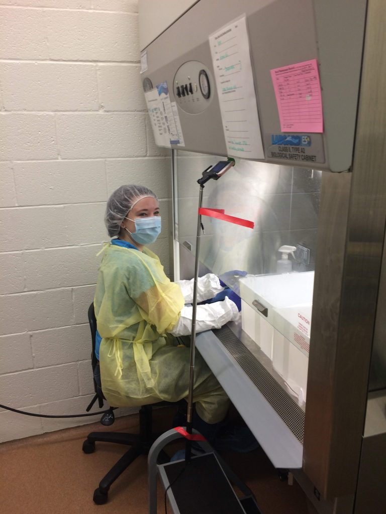
Will work when needed: in the lab on a weekend with a cast on my foot (visible on the left leg).

Being at the Oregon National Primate Center, Sweta’s model organism is the “Rhesus macaque” monkey. These monkeys have a genome ~97.5% similar to humans, meaning that the work she does is very relevant and translatable to humans. Working with the monkeys also means that her research is variable depending on the day. The monkeys will sometimes undergo treatments similar to those done in human IVF (in vitro fertilization) clinics, including surgeries to collect eggs for further research. After harvesting these eggs, they can be fertilized and the cells’ growth, division, and development can be monitored in a plate. When these experiments are not taking place, Sweta conducts various molecular biology experiments.

Sweta has become a true Pacific northwestener: hiking in rain with her husband through the Washington Park, Portland, OR. 


In India, Sweta completed her Bachelor’s degree at Dr. D. Y. Patil university in biotechnology and her first Master’s at SRM Institute of Science and Technology. During this time, Sweta happened to have several of family and friends undergoing IVF treatments and also worked in a fertility clinic for a time, bringing her attention to scientific needs within this field. Sweta then completed a second Master’s in Biological Sciences with a fellowship from the California Institute for Regenerative Medicine, and fell in love with fertility-related research during an internship at Stanford where she worked on embryo development. Her passion for this field of research led her to OHSU.

In addition to a being an accomplished researcher, Sweta is also an accomplished Indian Classical Dancer! She teaches bharatanatyam dance classes out of her home and travels around the US to perform. Long term, she hopes to continue research and also run a dance company.

Sweta will be presenting a piece on “depression” to work towards mental health awareness October 25th through 27th. The piece will be in Bharatanatyam and presented as a part of the 12th residency performance at N.E.W. 

Sweta writes her own blog posts about her journey through grad school which can be found here: 

  1. https://blogs.ohsu.edu/studentspeak/2017/09/11/it-is-possible-to-make-sad-not-even-seasonal/
  2. https://blogs.ohsu.edu/studentspeak/2018/07/24/phd-is-more-than-your-research/
  3. https://blogs.ohsu.edu/studentspeak/2019/04/18/never-give-up-there-is-a-bright-day-out-there-drudnischay/

To hear more about Sweta’s graduate work, personal struggles, and classical Indian dance moves, tune in on Sunday, October 20th at 7 PM on KBVR 88.7 FM, live stream the show at http://www.orangemedianetwork.com/kbvr_fm/, or download our podcast on iTunes!

The bacteria living inside us and what they have to say about autism

Trillions of bacterial cells are living within us and they’re controlling your brain activity.

Grace Deitzler is a 2nd year PhD student in microbiology working in Dr. Maude David’s lab on the gut-microbiome and its relation to autism spectrum disorder.

The gut-microbiome is the total population of bacteria living within our digestive tract. These bacteria are critical for digestive health, but also for our immune system and mental health. For example, we harbor bacteria capable of digesting plant fibres we otherwise could not digest. And if you’ve been told that probiotics are good for you, that’s because probiotics can change the gut microbiome in a positive way, allowing for increased bacterial diversity associate with improved health. These bacteria communicate with each other through chemical signaling but also communicate with us. Tryptophan, for example, is an amino acid produced through bacteria metabolism and is a precursor for serotonin, a brain-signaling chemical which causes feelings of happiness.

When the gut communicates with the brain, we call this, the “gut-brain axis”. Grace’s work narrows in on the gut-brain axis and more specifically, how one bacterial species in particular impacts autism spectrum disorder. To further complicate things, the gut-microbiome helps to regulate estrogen levels, and we also know that autism is a disorder found primarily in biological males. Which leads Grace to one of her biggest questions: are the bacteria involved in endocrine system regulation in women, also that responsible for this variation we see. Grace uses a mouse model to elucidate underlying mechanisms at play.

Step one is to feed the mice bacteria that have been found in elevated amounts in people with autism spectrum disorder than in neurotypical peers. These bacteria will colonize in the gut, and mice will go through several behavioral tests to determine if they are exhibiting more behaviors associated with autism. Grace performs three types of tests with the mice: one to test inclination to form repetitive behaviors, one to test anxiety, and one to test social behaviors. One test is a marble-burying test, in which a mouse more inclined to form repetitive behaviors will bury more marbles.

After behavioral testing is complete, the mice are sacrificed and different regions of the gut are taken to look for presence of bacterium. Tissues taken from the mice are used to look for transcriptional markers. The transcriptome is collected for both the mouse and the bacteria present, or the sum total of all genes that are read and converted to RNA. RNA are able to be isolated and sequenced using distinctive markers such as a “poly-A tail”. After this data is collected, Grace can finally move to the computational side of her work which involves combining biological and biochemical data with her behavioral studies.

In addition to her work on autism spectrum disorder, Grace also has a side project working in a honey bee lab, looking at the gut microbiome of honey bees in response to probiotics on the market for beekeepers. But Grace is one very busy bee herself because in addition to her lab work, she’s also involved with an art-science club called “seminarium”. The club is filled with scientists interested in art and artists interested in science. Grace is a painter primarily but is also working on ink illustration. The focus of this group is that art and science are complimentary, not at odds. The group has produced some collaborative projects, including a performance for a lab studying a parasite that effects salmon. The group put together a collage of interpretations of the parasites and had a performance in which one member played piano while someone else drew the parasite live.

Grace moved to Oregon from St. Louis Missouri. She completed her undergraduate degree in biological sciences with minors in chemistry and psychology at a small engineering college, Missouri University of Science and Technology, where she was a radio DJ! Grace first became involved in research during a summer internship in a microbiology lab at Washington University. There she studied the vaginal microbiome and how it effects pregnancy outcomes. Grace went back to this lab for the next couple summers and produced 4 publications! Ultimately, Grace graduated college early after they offered her a full time research position where she worked for a year and a half as a research tech. Through this experience, Grace came to realize that medical school was not her path, canceled her scheduled MCAT and signed up for GRE. Grace looked for schools in the PNW because she knew she wanted to live there, got an interview at OSU, loved it, and here we are!

Join us at 7 pm on Sunday, August 11th, 2019, to hear more about Grace’s research and her journey to OSU. Stream the show live on KBVR Corvallis 88.7FM or check out the episode as a podcast after a few weeks.

This time, it actually is rocket science: computational tools for modeling combustion

A.J. Fillo is in his final year of his PhD in Mechanical Engineering in the School of Mechanical, Industrial, and Manufacturing Engineering, within the College of Engineering. Working with Dr. Kyle Niemeyer. A.J. is studying combustion, or how things burn; specifically, A.J. is working to better understand how the microscopic motion of molecules impacts the type of combustion that we use in jet engines.

From A.J.’s masters work, and an photo-art series A.J. did on combustion, Turbulent, premixed jet fuel air Bunsen burner with a fuel rich jet fuel air flame. Fuel is commercially available ‘Jet-A.’ Photo shot at 1/8000 second shutter speed and aperture of f/2.8

            To understand combustion, first it’s helpful to understand energy.  If you drop a ball at the top of a hill, it will roll to the bottom, if you place a tea bag into a hot glass of water, the flavors will move through the water until you have tea. Both of these processes take something from its high energy state, to a more stable lower energy state. In our tea cup, molecular diffusion is what moves that energy around. Diffusion is the process of molecules becoming evenly dispersed by moving from high to low concentration and happens at very small scales, and affects everything around us including the combustion that we use in jet engines.

Diffusion is only part of the story though.  In fluid mechanics, the study of how gasses and liquids move around, diffusion controls the smallest aspects of motion but what processes control motion on a larger scale? To answer that A.J. used the example of an airplane wing. In physics class, many of us have seen a drawing or a demonstration of an airplane wing with smooth streaks of air flow over it, we call those smooth air streaks streamlines.  These smooth streamlines represent something called laminar flow, which is very smooth and predictable, but fluid flows are rarely predictable, usually they are swirly, changing, and chaotic.  These chaotic flows are called turbulence and exist all around us, they cause planes to bounce around when we fly through rough air, they drive the little vortex tornado the forms when our sink drains, and they can even impact the motion, structure, and chemistry of a jet fuel flame.

2D slice of a 3D simulation results for a turbulent, premixed, n-heptane air flame looking at flame temperature. Flow is from left to right.

Both turbulence and diffusion work to move energy around in combustion, but we don’t yet have a firm understanding of how these two different processes interact to control the combustion we use to propel us through the air.

Turns out, flames are hard to study because as you can imagine, anything you would use to measure a flame, does not want to be in a flame; measurement tools like thermocouples and pressure transducers can melt, or even combust themselves.  But there is another tool at our disposal.  We can use super computers to simulate how combustion is happening in jet engines and even use it to study how turbulence and diffusion interact, or how molecules are moving around during combustion.

From A.J.’s masters work, and an photo-art series A.J. did on combustion, Turbulent, premixed jet fuel air Bunsen burner with a fuel lean jet fuel air flame. Fuel is commercially available ‘Jet-A.’ Photo shot at 1/8000 second shutter speed and aperture of f/2.8

A.J.’s research focuses on developing computational tools to look at these effects. The sum total of reactions happening during jet fuel combustion are large and complex, meaning that the equations are not easy to solve, and trying to do so can take thousands of computer cores for several days. By developing a more efficient computer algorithm to look at these reactions we can make these simulations faster, more efficient, and less expensive.

In reality, Jet fuel is a mixture of hundreds of different chemicals, so to simplify things, A.J. uses fuels like hydrogen (H2), n-heptane (H3C(CH2)5CH3), and toluene (C6H5CH3) as representative fuels. Although a single, simpler compound, even as simple as just hydrogen, has hundreds of chemical reactions and dozens of different radical molecules that form during its combustion. To get around the limitation of computer memory and speed up how quickly his simulations run, A.J. created an algorithm to optimize how the computer handles the math to make sure things run as smoothly as possible.  You can think of it a bit like going to the DMV, usually the line takes forever because people are rarely ready with their paper work in hand when they get to the front of the line, instead people must get out of line, get more paper work, and start over.  Using this analogy, A.J.’s algorithm works to make sure everyone in line arrive with their paper work completed, ready to hand off, and let the next person through. This reduces dramatically reduced the amount of computer memory needed to solve these combustion simulations and speeds up the math.

3D simulation results for a turbulent, premixed, hydrogen air flame looking at peak flame temperature colored by chemical composition. Flow is from back to front

A.J. became interested in mechanical engineering because of his love of magic. A.J. started his academic journey at the University of Missouri Columbia as a journalism major but transferred to OSU for the engineering program. A.J. has always loved performing, which is why science outreach has been such a large part of his graduate school experience. Partnered with the Corvallis Public Library, A.J. hosts LIB LAB, a hands-on multimedia educational YouTube series focused on STEAM (science, technology, engineering, arts, and mathematics) education, which he previously talked about on our GRADx event.

A.J. standing with the Oregon State University Drumline in OSU’s Reser Stadium while filming an episode of his YouTube show LIB LAB about vortex smoke rings.

To find out more about A.J.s research, outreach, and journey to grad school, join us on Sunday, May 12 at 7 PM on KBVR Corvallis 88.7 FM or stream live.


3D Modeling Rock Shape: Archeological Research of the Earliest North Americans

At age 17, like a lot of teenagers, Samuel Burns wanted to go to college. Unlike most college-bound 17-year-olds however, Samuel didn’t have a high school degree. Today, Samuel is a first-year master’s student in Applied Anthropology, within the School of Language, Culture, and Society, and the Department of Anthropology. Also, this is his second master’s degree.

Samuel in the field in the Allegheny National Forest, Pennsylvania. Photo by Samuel Burns.

Samuel works with Dr. Loren Davis to investigate the earliest archeological sites in North America, and there are two big questions to answer: when did humans first arrive in North America, and by what route did the earliest humans arrive? Traditionally, humans are thought to have entered North America through the Rocky Mountains, but more recent evidence suggests that maritime cultures may have arrived first, finding North America via the ocean. The oldest fish hooks in North America are somewhere between ~11,300 to 10,700 years years old and were discovered off the coast of Baja California, Mexico on Cedros Island.

Cedros Island is just one of two archeological sites of interest for Samuel’s research group, and while he has been to Cedros to conduct fieldwork, Samuel’s work focuses on artifacts from one pit in the second site: Cooper’s Ferry in Cottonwood, Idaho, near the Salmon River. From Cooper’s Ferry, seemingly interesting artifacts are brought back to the lab where they are sorted, confirmed to be artifacts, and studied.

L-R: Loren White (OSU), Steve Jenevein (Oregon State Parks), and Samuel Burns on board the flight from Cedros Island, Baja California, Mexico after a successful field session in January, 2019. Photo by Samuel Burns.

Samuel is able to take the artifacts, make 3D scans of the object, and input this information into a computational program. The computer converts the 3D scans into mathematical shapes and 3D models. So instead of looking at a couple things by eye and estimating if artifacts are similar or different, the program can compare large sets of data with discreet numbers and make conclusions about whether or not two artifacts found in different places have similar shapes. This allows researchers to ask questions about tool development over time and place.

To make 3D images, a laser scanner has been used in the past, but this is both expensive and large, so new methods are actively being developed for this purpose. One option is a structured light scanner, which has a light shining through multiple holes. To use a structured light scanner, you place your artifact on a patterned background and take lots of photos at many angles, producing a large amount of data to feed the computer program. Another easier option for 3D modeling is photogrammetry, which only requires a camera and a computer, even just a phone camera will work. This soft ware used is called “GLiMR” (GIS-based Lithic Morphometric Research) and is based on GIS software for modeling geographical landscapes, and the automation and ease of such a program enables archeologists to spend less time collecting numbers and more time assessing these numbers through statistical analyses and asking interesting questions.

Samuel’s crew lining up to conduct a systematic surface survey near Paulina, Oregon. Photo by Samuel Burns.

When you think about ancient North American stone artifacts, megafauna hunting tools like arrow heads and spears come to mind. However, in both the Cedros and Cooper’s Ferry sites, simpler tools are being found that suggest early North Americans exploited a wide range of resources and had a broad-spectrum diet. For example, artifacts found include shell or stone tools for processing fiber to making fishing line.

Samuel using a digital total station to take measurements at a Medieval Christian period site at el Kurru, Northern State, Sudan. Photo by Walter De Winter.

Growing up, Samuel never went to school and wasn’t homeschooled, but always loved history. He lived in an 1850s farmhouse, and spent his childhood going through old objects from his backyard, left behind over the past 100+ years. At age 17, realizing he wanted to go to college but not having the traditional requirements, Samuel applied to a University in Jerusalem and got in. After spending a year there, he ran out of money, and spent next few years working and moving around the world, including in South Korea and Israel. Eventually, he returned to the US and jumped back into school at a community college in Michigan and ultimately transferred to the University of Michigan, where he focused on ancient cultures and language of middle east.

Field camp near Colt, Arkansas, home for 6 months in 2016-2017. Photo by Samuel Burns.

Samuel graduated from UM in 2010 and then got a master’s degree at the University of Cambridge in the United Kingdom, focusing on Egyptian studies. This first master’s centered around Syria and unfortunately, this research project was not able to be pursued further, so Samuel spent the next five years working in cultural resource management in the US. Through this job, he was able to travel around the US and soon became interested in North American archeological research. Samuel had a strong liberal arts background but, wanting to expand his earth science knowledge, came to Oregon State.

Eventually, Samuel wants to obtain a PhD and work in academia, continuing to formulate and direct research projects.

To hear more about Samuel’s path to OSU and experiences in archeological research, tune in Sunday, February 16th at 7 PM on KBVR 88.7 FM, live stream the show at http://www.orangemedianetwork.com/kbvr_fm/, or download our
podcast on iTunes!
 

Davis, L. G., Bean, D. W., Nyers, A. J., & Brauner, D. R. (2015). GLiMR: A GIS-Based Method for the Geometric Morphometric Analysis of Artifacts. Lithic Technology, 40(3), 199–217.
Des Lauriers, M. R., Davis, L. G., Turnbull, J., Southon, J. R., & Taylor, R. E. (2017). The Earliest Shell Fishhooks from the Americas Reveal Fishing Technology of Pleistocene Maritime Foragers. American Antiquity, 82(3), 498–516.

Kayaks and Computers: the Gray Whale Research Essentials

Throughout the year, looking out from the Oregon coast, you can often spot gray whales with the naked eye. Behind the magic and mystery of these massive creatures are teams of researchers tracking their migration and studying their diet.

Lisa Hildebrand is a 1st year Master’s student in Wildlife Science working with Dr. Leigh Torres within the College of Agriculture. Lisa studies geospatial ecology of marine megafauna, meaning that her research focuses on the feeding and movement through time and space of sea creatures larger than most fish, including large sea birds, seals, dolphins, and of course, the gray whales. To study such large animals in the ocean, Lisa manages a team that combines diverse technologies coupled with fine scale foraging ecology.

Gray whales feed on very small zooplankton suspended in shallow water. The whales don’t have teeth but instead have rows of baleen which look like a thick brush and act as a filter for water and sediment while letting in large quantities of zooplankton. In July and August, Lisa and her team of 4-5 people go out to Port Orford, Oregon. The team splits into two groups: a cliff team and a kayak team. From a cliff above their 1km2 sampling site, theodolites and computational programs are used to track whales by height and GPS location. Once a whale is spotted, team members kayak to this location and take water samples for analysis of zooplankton density, caloric content, species, and microplastic quantity. Lisa has taken over this ongoing project from a previous Master’s student, Florence Sullivan, and has data on the same research site and whales going back to 2015.

This research project provides opportunities for both undergraduates and high school level students to obtain first-hand field research experience. The students involved are able to take what they’ve heard in a classroom and apply it outdoors. In particular, Lisa is passionate about getting the students in the local Oregon coastal community involved in research on the whales that bring many tourists to their area.

To study the large gray whales, Lisa spends most of her time studying the small zooplankton that they eat. Zooplankton hide under kelp and it turns out, can be separated by populations that are pregnant, or varied in age or species. Gray whales may show preference for some feeding sites and/or types of zooplankton. Why do we care what a gray whale’s dietary preferences are? Plastic use and plastic pollution are rampant. Much of our plastic ends up in the oceans and photodegrade into microplastics small enough to be consumed by zooplankton. Since gray whales are the top predator for zooplankton and eat large qualities, these microplastics accumulate. Microplastic presence may differ between regions and species of zooplankton, which may relate back to whale preferences and migratory patterns. On the Oregon coastline, microplastic profiles of zooplankton have not yet been studied. As humans are also consuming large quantities of seafood, it is important to understand how microplastics are accumulating in these areas.

Lisa is from Germany and grew up in Vietnam and Singapore, but she was first inspired to pursue marine animal research as a career after a family trip to Svalbard, Norway during high school. Before obtaining her undergraduate degree in Marine Zoology from Newcastle University in England, Lisa took two years off from schooling and completed two internships: one with bottlenose dolphin sanctuary research institute in Italy and Spain, and one at a seal research facility in Germany. Now that she’s settled in Oregon for now, Lisa is enjoying the nature and in her free time loves hiking and skiing.

To learn more you can check out GEMM Lab website , the GEMM Lab blog and Lisa’s Twitter, @lisahildy95

To hear more about Lisa’s research, tune in Sunday, January 20th at 7 PM on KBVR 88.7 FM, live stream the show at http://www.orangemedianetwork.com/kbvr_fm/, or download our podcast on iTunes!

Treating the Cancer Treatment: an Investigation into a Chemotherapy drug’s Toxic Product

One of the most difficult hurdles in cancer treatment development is designing a drug that can distinguish between a person’s healthy cells and cancer cells. Cancerous cells take advantage of the body’s already present machinery and biochemical processes, so when we target these processes to kill cancer cells, normal, healthy cells are also destroyed directly or through downstream effects of the drug. The trick to cancer treatment then is to design a drug that kills cancer cells faster than it harms healthy cells. To this end, efforts are being made to understand the finer details that differentiate the anti-cancer effects of a drug from its harmful effects on the individual. This is where the research of Dan Breysse comes in.

Dan a third-year master’s student working with Dr. Gary Merrill in the department of Biochemistry and Biophysics. Dan’s research focuses on a common chemotherapy drug, doxorubicin. Doxorubicin has been researched and prescribed for about 40 years and has been used as a template over the years for many other new drug derivatives. This ubiquitous drug can treat many types of cancer but the amount that can be administered is limited by its toxic effect on the individual. Nicknamed “the red death,” doxorubicin is digested and ultimately converted to doxorubicinol, which in high doses can cause severe and fatal heart problems. However, hope lies in the knowledge that doxorubicinol generation is not related to the drug’s ability to kill cancer cells. These mechanisms appear to be separate, meaning that there is potential to prevent the heart problems, while keeping the anti-cancer process active.

Cancer cells replicate and build more cellular machinery at a much faster rate than the majority of healthy cells. Doxorubicin is more toxic to fast-replicating cancer cells because its mechanism involves attacking the cells at the DNA level. Dividing cells need to copy DNA, so this aspect of doxorubicin harms dividing cells faster than non-dividing cells. It is common for chemotherapy drugs to target processes more detrimental to rapidly dividing cells which is why hair loss is often associated with cancer treatment.

Separately, doxorubicin’s heart toxicity appears to be regulated at the protein level rather than at the DNA level. Doxorubicin is converted into doxorubicinol by an unknown enzyme or group of enzymes. Enzymes are specialized proteins in the cell that help speed up reactions, and if this enzyme is blocked, the reaction won’t occur. For example, an enzyme called “lactase” is used to break down the sugar lactose, found in milk. Lactose intolerance originates from a deficiency in the lactase enzyme. During his time at OSU, Dan has been working to find the enzyme or enzymes turning doxorubicin into doxorubicinol and to understand this chemical reaction more clearly. Past research has identified several potential enzymes, one of which being Carbonyl reductase 1 (CBR1).

Doxorubicin is converted to doxorubicinol with the addition of a single hydrogen atom.

While at OSU, Dan has ruled out other potential enzymes but has shown that when CBR1 is removed, generation of doxorubicinol is decreased but not completely eliminated, suggesting that it is one of several enzymes involved. In the lab, Dan extracts CBR1 from mouse livers, and measures its ability to produce doxorubicinol by measuring the amount of energy source consumed to carry out the process. To extract and study CBR1, Dan uses a process called “immunoclearing,” which takes advantage of the mammal’s natural immune system. Rabbits are essentially vaccinated with the enzyme of interest, in this case, with CBR1. The rabbit’s immune system recognizes that something foreign has been injected and the system creates CBR1-specific antibodies which can recognize and bind to CBR1. These antibodies are collected from the rabbits and are then used by Dan and other researchers to bind to and purify CBR1 from several fragments of mouse livers.

Prior to his time at OSU, Dan obtained a B.S. in Physics with a concentration in Biophysics from James Madison University where he also played the French horn. Realizing he loved to learn about the biological sector of science but not wanting to completely abandon physics, Dan applied to master’s programs specific to biophysics. Ultimately, Dan hopes to go to medical school. During his time at OSU, he has balanced studying for the MCAT, teaching responsibilities, course loads, research, applying to medical schools, and still finds time to play music and occasionally sing a karaoke song or two.

To hear more about Dan’s research, tune in Sunday, December 16th at 7 PM on KBVR 88.7 FM, live stream the show at http://www.orangemedianetwork.com/kbvr_fm/, or download our podcast on iTunes!

Finding cancer with sound: the development of nanoparticles to deliver light-to-sound converting agents

“Here I am!” -Cancer

Wouldn’t it be nice if cancer could simply yell out to let us know where it is, and how much of it is there? Anna St. Lorenz, a 4th year PhD student in the College of Pharmacy, is working on just that.

Anna volunteering at Brain Day at OMSI science museum.

Anna’s path to scientific research began when she was 8 years old, on a farm, with some chickens and a candle-lit microscope. Anna spent much of her childhood becoming familiar with the local ecology, as well as the Mendelian laws of genetic inheritance that applied to her family’s chicken breeding. However, her first taste of research was in Death Valley. With funding provided through Smith College associated religious programs, Anna studied arsenic-eating-microbes, but thanks to some giant spiders and allergies, Anna decided field research wasn’t for her and moved to a hospital setting.

In college, Anna’s scientific education expanded further through multiple internships and unique educational opportunities at Novartis Pharmaceuticals, Dana-Farber Cancer Institute and OHSU. Anna obtained a B.A. in Biology with a minor in Neuroscience from Smith College. Receiving a B.A. rather than a B.S. meant that Anna’s science education was interdisciplinary, and incorporated disciplines such as history and the fine arts. Anna’s love of the arts still persists as she frequently paints and creates “bioart,” which she uses as a means to inform and involve the community on her scientific endeavors. She commonly uses her work with her husband, Grey St. Lorenz, in presentations and has previously collaborated with artists in upstate New York for work on display at local universities. 

Bioart by Anna. Nanoparticles taken up by an endosome, that then create a pore in the endosome’s membrane to release their payload. It is done in the style of Starry Night and the nanoparticle’s payload matches up with the stars.

After completing her undergraduate degree, Anna received a Master’s in Biomedical Engineering from Rensselaer Polytechnic Institute. While finishing up her Master’s degree, Anna moved to Boston and started working at MIT as a nanoparticle research technician within the Langer Lab. It was at MIT that she learned about a new nanoparticle-specific program being implemented in the OSU College of Pharmacy. This program is now about four years new and Anna has been at the front line of pioneering this program for future graduate students. In addition to navigating a new program and coping with the regular difficulties of being a graduate student, this OSU nanoparticle program is actually based at the Oregon Health & Science University (OHSU) in Portland. Although challenging at times, as a graduate student researching cancer therapeutic technology, OHSU is great place to be.

Anna and the Taratula group.

In this program, Anna works with the Taratula group on ovarian cancer diagnosis. As a disease that is traditionally hard to detect at early stages, it is often only after the cancer has spread to other areas of the body in later stages that diagnosis is able to be made. This metastasis results in a worse prognosis and decreased survival rates. To this end, Anna and other researchers and medical professionals are developing nanoparticles to deliver various iterations of imaging agents. Anna’s role in this process is to design more specific nanoparticles to carry various agents through the bloodstream and allow for specific staining of cancerous tissue.

Bioart by Anna and Grey St. Lorenz demonstrating a nanoparticle (blue) encapsulating a compound (red) and adorned with targeting antibodies (green).

Have you ever used facewash with textured particles in it?  Nanoparticles are 1/1000th of that size and are used to envelop or otherwise transport compounds throughout the body and deliver them to more specified regions. This technology can be applied to a variety of compounds to enhance their delivery needs. Solubility issues, tissue or disease specificity, PH, heat, and enzyme specific release are all areas that nanoparticle science delves into to address patient care. So now, the imaging agent, inside of its tiny carrier, can circulate through the body and find the cancerous tissue it’s designed to target.

As tumors are characteristically disorganized tissue whose unregulated growth demands increased nutrients, they develop a leaky vasculature  which makes it easier for molecules to permeate the tissue. Once the nanoparticle reaches the tumor, it is able to take advantage of the enhanced permeability of tumors to infiltrate and label the cancer cells. An important characteristic of the works is that the compounds use near-infrared (near-IR) light, which can be administered to excite the delivered agents in a spectral range that is largely unaffected by organic tissue. These agents were specifically screened for their ability to convert this light to acoustic/sounds waves that are detectable by ultrasound imaging.  This process allows for an enhanced detection and characterization of ovarian cancer – opening the door for effective screening and improved monitoring of this devastating disease.

Join us Sunday November 4th at 7PM on 88.7FM, or listen live, to learn more about Anna’s exciting journey to graduate school, bioart, sound-making cancer, and nanoparticles.